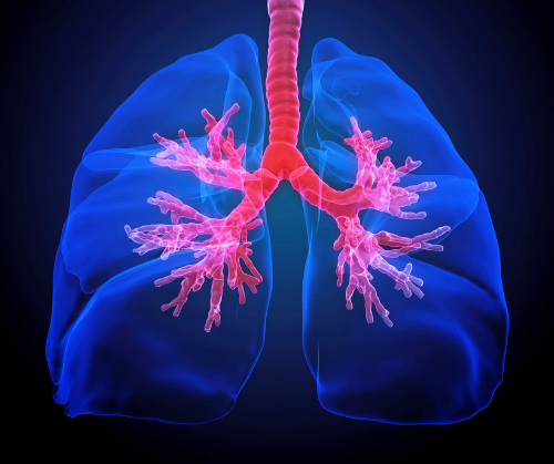
Bronchiectasis is a condition that affects hundreds of thousands of Americans each year. While COPD is the most common cause of bronchiectasis, individuals with genetic conditions like cystic fibrosis and alpha-1 antitrypsin deficiency account for approximately 10% of cases.
Bronchiectasis (braang·kee·ek·tuh·suhs) means “dilated airways.” When large or dilated airways in any part of the lung have trouble clearing mucus correctly, the effect is a chronic cycle of mucus buildup that attracts bacteria. As bacteria collect and multiply, infection in the airways leads to inflammation. Every new flare-up (or exacerbation) causes additional damage to the airways. The problem then becomes a chronic cough. The repeated infections are treatable, but each flare-up makes it harder to clear mucus from the airways and results in irreversible damage. The more widespread the damage, the more serious the disease. Therefore, bronchiectasis is a treatable condition with no cure.
Bronchiectasis was first defined by French physician René-Théophile-Hyacinthe Laennec in the 19th century. While working with patients with chronic development of phlegm, he observed: “The affectation of the bronchiectasis is always produced by some other disease attended by long, violent, and often repeated fits of coughing.”
In the United States, bronchiectasis diagnoses are growing at a rate of 8% per year, with more women than men diagnosed. It is an expensive disease with estimated treatment costs exceeding $630 million annually due to the need for repeated treatments and antibiotics.
The most common causes of bronchiectasis are chronic obstructive pulmonary disease (52%), acute bronchitis (38%), post-inflammatory fibrosis (14%), and genetic disorders (10%) like Alpha-1 and cystic fibrosis.
Cystic fibrosis is possibly the most recognized cause of bronchiectasis and affects approximately 30,000 children and adults in the US alone. There are also individuals with bronchiectasis for no obvious cause. This is called idiopathic bronchiectasis. However, when bronchiectasis is due to Alpha-1, this is called Alpha-1-associated bronchiectasis.
A study conducted in 2007 revealed the connection between Alpha-1 and bronchiectasis. In the study population, the prevalence of Alpha-1 ZZ patients with bronchiectasis was 27%. The age range for affected individuals in the study was 56-74 years. It is thought that neutrophils, a type of white blood cell, have a key role in the development of bronchiectasis in Alpha-1 patients.
Common symptoms associated with bronchiectasis include: coughing up mucus, coughing up blood or blood mixed with mucus, whistling or wheezing sounds, shortness of breath, unexpected weight loss, and chest pain during flare-ups.
Radiology plays a key role in the diagnosis of bronchiectasis; in fact, the diagnosis cannot be made without a chest computed tomography (CT) scan. Findings on a bronchiectasis positive scan show thick and dilated airway walls, mucus plugging, and tree-in-bud opacities. In some cases, blood tests may be required to diagnose Alpha-1 or cystic fibrosis, as well, and sputum cultures may be taken to find out if there are infections that require treatment. Pulmonary function tests can help determine how well the lungs are working while a bronchoscopy, which is a way to see inside the lungs, can determine if there are mucus blockages and remove them. During this procedure, mucus can be obtained for further cultures.
Treating bronchiectasis is individualized based on severity and the cause. The primary goal for all individuals is to prevent inflammation and flare-ups and ensure airway clearance.
Hand-held devices, electronic chest clappers, vest and chest physical therapy, and aerobic exercise are popular measures for helping to reduce and prevent bronchiectasis-associated exacerbations. Mucus thinners make mucus easier to cough up. Inhaled bronchodilators, inhaled corticosteroids, and nebulized hypertonic saline may be used to loosen airway mucus for easier clearance. Antibiotics can be given as pills or as inhaled drugs to treat bacterial infections of the airways. Oral macrolide antibiotics, such as Azithromycin, Erythromycin, and Clarithromycin can treat infections and have anti-inflammatory properties at the same time.
The outlook for people with bronchiectasis is much improved after the condition is diagnosed with a chest CT scan. Controlling exacerbations is the key to a better quality of life and a normal lifespan.
Sources:
Milliron, Bethany, Henry, Travis S., Veeraraghavan, Srihari and Little, Brent P., Bronchiectasis: Mechanisms and Imaging Clues of Associated Common and Uncommon Diseases RadioGraphics 2015 35:4, 1011-1030.
Parr DG, Guest PG, Reynolds JH, Dowson LJ, Stockley RA. Prevalence and impact of bronchiectasis in alpha1-antitrypsin deficiency. Am J Respir Crit Care Med. 2007 Dec 15;176(12):1215-21. doi: 10.1164/rccm.200703-489OC. Epub 2007 Sep 13. PMID: 17872489.
Weycker, D., Hansen, G. L., & Seifer, F. D. (2017). Prevalence and incidence of noncystic fibrosis bronchiectasis among US adults in 2013. Chronic respiratory disease, 14(4), 377–384. https://doi.org/10.1177/1479972317709649.
